But We In-Cyst: Identifying, Diagnosing + Treating Canine Cysts
Remember, this is an educational resource, not a guide for diagnosis.
Pictures are helpful, but they may not tell the whole dermatological story. We always recommend consulting with your vet for any of your dog's health concerns.
Want a Reusable 20% Off Coupon?
Sign up for our scarce + savvy emails and get a discount code that you can apply all season long!
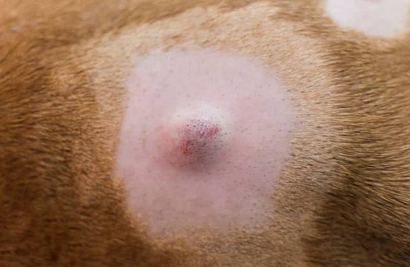
What Are Cysts?
Cysts are hollow spaces or sacs within tissue that contain liquid, semi-liquid, or solid materials. These materials could be secretions that occur in the body naturally, such as sweat or sebum, or atypical breakdown products such as dead tissue cells, blood, or keratin.
Cysts tend to be small (0.5cm - 5cm), round in shape, and well-defined. They can occur anywhere on a dog's body, though some areas may be more susceptible than others, depending on the type of cyst.
Photo: Sebaceous cyst, via Kingsdale Animal Hospital
Types of Cysts on Dogs
There are several different kinds of cysts, nearly all of which are benign and non-cancerous. That said, it is always a good idea to get cysts or suspicious bumps on your dog examined by your vet to ensure that they are not cancerous or a sign of an underlying disease.
True Cysts
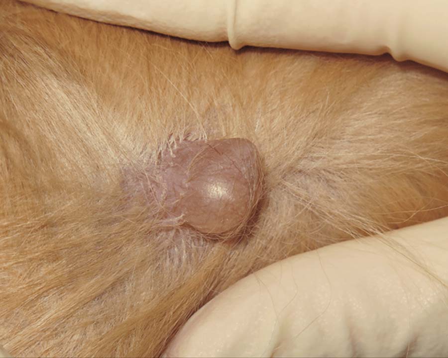
True cysts have a membrane (called an epithelial or secretory lining) on their inner surface that produces secretions. These cysts typically arise due to a blocked passageway, resulting in the area filling with excess fluid like a balloon.
True cysts often form in glands (especially sweat glands, as on the eyelids) where fluids are produced, appearing slightly translucent with a bluish or dark color. They may appear in numbers around the eyes and ears, and secrete a yellowish substance.
True cysts can be surgically removed, but the lining must be completely taken out or the cyst may return.
Photo: Canine cyst, via Today's Veterinary Nurse
False Cysts
False cysts don't have an epithelial lining and are better referred to as a "fluid-filled mass." They are often caused by hemorrhage or trauma - the latter being more common in dogs.
The trauma - for example, from rough play or collision with something sharp - kills the tissue and creates a pocket that fills with blood and dead tissue that becomes liquified. Because of this, false cysts typically appear dark-colored. They can sometimes appear due to a reaction to an injection.
Dermoid Cysts
Dermoid cysts are quite rare, forming while in the mother's womb. These cysts occur because the outer layer of skin (epidermis) does not close properly. Rhodesian Ridgebacks and Kerry Blue Terriers are breeds that tend to have these cysts.
Follicular Cysts
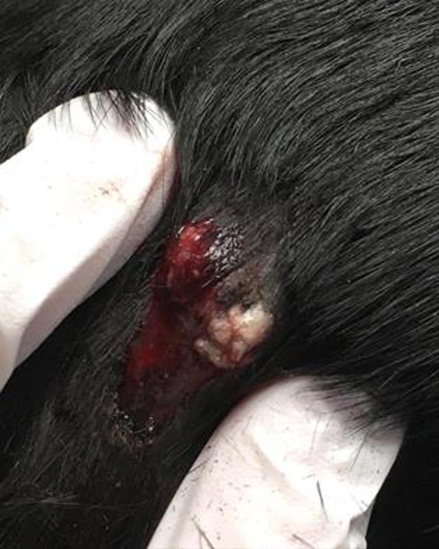
Follicular cysts represent a majority of cysts found on dogs. These cysts, also called epidermoid cysts, form when a hair follicle becomes inflamed, blocked, or damaged due to localized injury, pressure (especially at pressure points, such as the elbow), UV damage, or inactivity (seen mostly in hairless breeds).
Follicular cysts look like round hard tissue masses (nodules) on or underneath the skin and may contain fluid or a thick, cheese-like substance (keratin). They can become itchy or painful as they grow in size.
Follicular cysts are prone to bacterial infection (pyoderma), and the discharge will then take on a foul odor. They tend to appear on the head, neck, or abdomen, but can show up anywhere on a dog.
Photo: Ruptured follicular cyst on dog, by Dr. Roberta Riedi, DVM, via the Veterinary Information Network
Sebaceous Cysts
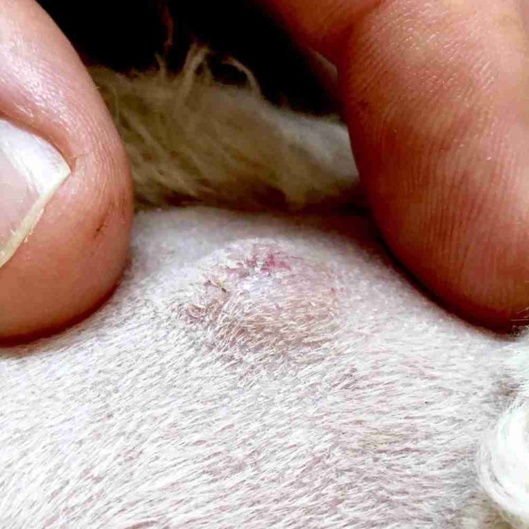
Sebaceous cysts are very similar to follicular cysts, in that they affect a part of the hair follicle called the sebaceous gland. The sebaceous glands produce an oily substance called sebum, which helps lubricate and protect a dogs' skin and coat. When these glands become blocked - due to genetics, debris, trauma, or injury - the buildup of sebum forms a cyst.
Like follicular cysts, sebaceous cysts are common in dogs and prone to bacterial infection. They tend to appear a white or light blue color, and their discharge (built up sebum and/or other matter) will be a thick, gray or yellow-brownish color.
Sebaceous cysts are most commonly found on the head, neck, body, and hips. They don't tend to be itchy or painful unless they become damaged or occur in an area that experiences constant pressure or inhibits mobility.
Photo: Sebaceous cyst, via Walkerville Vet of Queensland
Interdigital Cysts
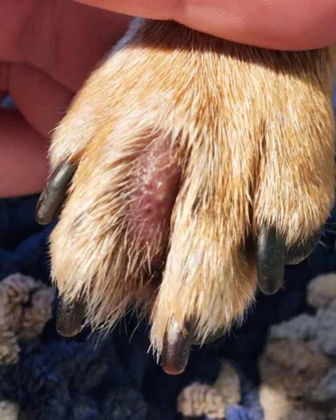
Interdigital cysts (furuncles) are follicular cysts that appear between a dog's toes - typically brought on by allergies, trauma, excessive pressure, or excess friction. Because these cysts occur in such a high-impact area, they tend to be irritating and itchy.
As a dog licks and chews at the cyst, they further damage the hair follicles and skin and add to the inflammation. Like other follicular cysts, interdigital cysts can develop into open sores and easily become infected.
Photo: Interdigital cyst on dog paw, by Jennifer Bailey, DVM; via the Whole Dog Journal.
Dog Breeds Prone to Cysts
Though any breed of dog can develop a cyst, there are some breeds that seem to be at an increased risk.
How Are Cysts Diagnosed in Dogs?
Canine cysts are chiefly diagnosed via two methods, a fine needle aspiration or a biopsy. Every examination will begin with a visual inspection and palpation - that is, examining the cyst by touch to assess its consistency, mobility, attachment to underlying tissue, and if there is any sort of sensitivity.
A fine needle aspiration (FNA) uses a small needle and syringe to draw a sample of cells from a cyst for analysis under a microscope. FNA is less expensive and invasive than a biopsy and helps to get an idea of the type of cells within the cyst.
A biopsy involves removing part or all of the cyst and having it examined by a pathologist under a microscope (also called histopathology). This method is more accurate, as it gives a more complete analysis of the cells within the cyst and determines whether the entire cyst was successfully removed. Histopathology is also useful in looking for cancer or other potential underlying diseases.
Treatment for Cysts on Dogs
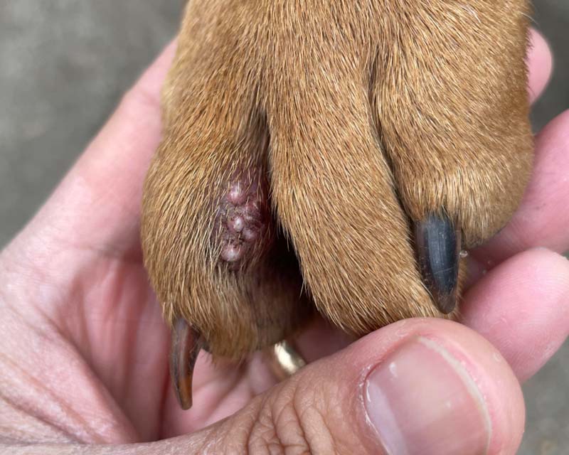
The most effective treatment of cysts in dogs is surgical removal. This ensures that the epithelial lining is removed so the cyst cannot reform. If there is an underlying cause(s) (disease, allergies, etc.), other treatments may be necessary.
False cysts due to trauma may resolve in time, or they can be drained by a veterinary clinic and treated with topical medications to prevent or counter infection.
Since cysts are typically benign, they do not always need to be removed immediately, but they should be monitored to check their growth rate.
It's very important that you do not manually squeeze a cyst in an attempt to burst or drain it, as the contents could leak down into the tissue and lead to further inflammation or infection. In short, you'll do more harm than good.
Caring for Canine Cysts at Home
You'll want to make sure your dog isn't licking, rubbing, scratching, or chewing at any cysts. This will bring on inflammation, further irritation, and increase the risk of infection.
If a cyst bursts or opens, you'll want to keep it clean and apply a topical ointment like Lavengel® to help it heal and protect it from bacteria.
If the cyst is surgically removed, Lavengel® can be used in the same way to protect the incision site. You will also want to be sure that your dog does not aggravate the site, so a bandage or e-collar (cone) may be necessary.
Photo: Enflamed hair follicles on dog paw which have possibly formed follicular cysts; via r/Lovefriedchicken on Reddit
Canine Cysts FAQs
Are my dog's cysts contagious?
No, cysts cannot be spread to other dogs, animals, or humans.
Do dog's cyst spread?
Cysts are isolated skin disruptions and do not spread across the skin.
They do not usually appear in numbers unless there is an underlying condition that is allowing for their growth. Certain hairless breeds, such as the Chinese Crested, may have multiple follicular cysts form due to inactivity of the hair follicles.
Warts, which can sometimes resemble cysts on dogs, do tend to appear in numbers, unlike cysts.
It should be noted that if you notice what appears to be a cyst that is growing noticeably quickly on your dog, it is more likely a tumor and should be checked out immediately.
Will my dog's cyst grow bigger?
Cysts grow slowly and can even remain the same size for years if left alone.
If you see a cyst on your dog that grows noticeably within a few months, it could a tumor and should be checked out immediately.
If a cyst is agitated or bursts, the contents can leak into the surrounding tissue and generate a lot of inflammation. On top of that, if the cyst is not completely removed, the bursted cyst can reform.
What's the difference between a cyst and a tumor?
Cysts are:
- Hollow spaces or sacs filled with fluid, semi-fluid, or solid material.
- Small, round raised bumps of a white, bluish, or translucent color with a well-defined border and can have a central pore or opening.
- Usually firm-soft (like rubber), yet pliable.
- Full of a discharge that is thick, yellow-brown or grayish.
Tumors are:
- Abnormal growths of tissue that can be benign or malignant.
- Not filled with another substance; no discharge
- Can be soft and pliable (lipomas, or fatty tumors) or harder to the touch and fixed to the deeper tissue.
- Usually larger and less defined than cysts.
Where do cysts generally occur on a dog?
Cysts can appear anywhere on a dog, but they are most commonly found on the head, neck, trunk, upper legs, and between the toes (interdigital) of dogs.
Are my dog's cysts cancerous, or can they become cancerous?
No, the vast majority of cysts are benign pockets of fluid trapped under the skin and cause no harm to your dog, nor do they become cancerous.
If anything, they are more likely to get infected by bacteria or fungi if they are ruptured.
Will my dog's cyst go away on its own?
In general, no, cysts will not go away on their own and must be surgically removed.
False cysts - pockets of blood, fluid, and/or dead tissue (no epithelial lining) that often result from trauma or injury - can recede and heal with time. However, it is best to get them checked by a veterinarian to avoid bacterial infection and an abscess forming.
Should I try to "pop" or drain my dog's cyst?
No. Attempting to burst a cyst may force the material within it to leak deeper into the skin and cause inflammation and pain. On top of that, if the cyst's inner lining is not removed, it will likely return.
Leave cysts to the professionals, folks.
Does Lavengel® help with dog cysts?
Lavengel® can be of help with false cysts or small abscesses, especially in countering bacterial infection or helping to reduce inflammation.
With true cysts, the epithelial lining of the cyst must be surgically removed to ensure the cyst does not return. Once removed, Lavengel® can help with incision lines and skin repair.
The Canine Skin Conditions Continue
-
Warts
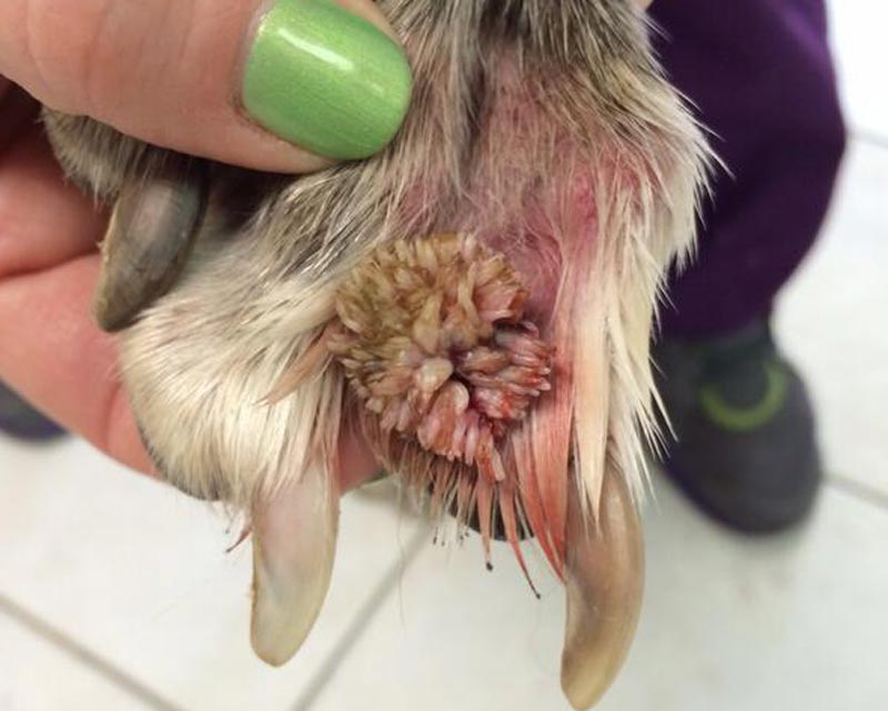
Wart's up with that?Warts, or papillomas, are benign tumors can occur between toes, on eyelids, ears, and genitals, and even inside your dog's mouth!
-
Dog Paw Licking
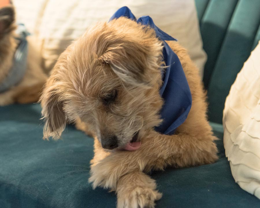
Dive in feet firstSo, why do dogs obsessively lick their paws like corn-chip-flavored popsicles? We've got answers, photos, and more.
-
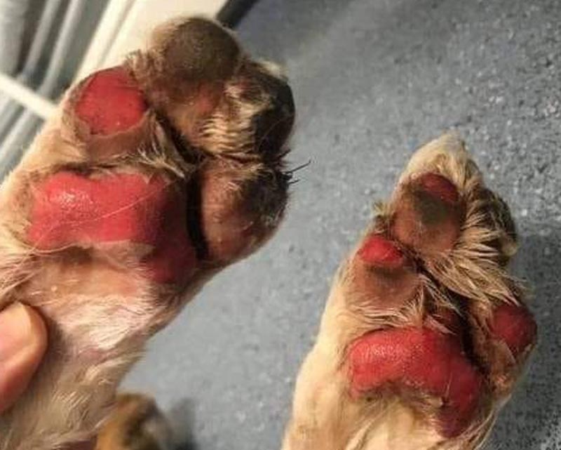
Burns
How to beat the heatWhile most dogs don't tend to receive thermal burns often, they do happen, and it is paramount to know what to do and why.
Collapsible content
Resources on canine cysts
- Today's Veterinary Nurse: What Is This Lump on My Pet?
- Walkerville Vet: Help! My Dog Has a Lump
- VCA Animal Hospitals: Cysts
- VCA Animal Hospitals: Interdigital Cysts in Dogs
- American Kennel Club: Types of Cysts on Dogs
- Kingsdale Animal Hospital: Sebaceous Cysts in Dogs
- Veterinary Information Network, Veterinary Partner: Follicular Cysts in Dogs
- PetMD: Lumps, Bumps, and Cysts on Dogs


